Imagine walking into a dental office where your x-ray results are instantly available, allowing for quicker diagnoses and treatment planning. This is the reality with digital x-ray technology, a game-changer in the dental industry. By replacing traditional film x-rays with advanced digital systems, dental practices can now offer enhanced patient care with greater efficiency and accuracy. This blog aims to shed light on the cutting-edge advancements in digital x-ray technology, examining how these innovations are reshaping the future of dental care and what they mean for both patients and practitioners.
In This Blog:
- Differences Between Traditional and Digital Dental X-Rays
- The Digital X-Ray Process: What to Expect
- Advancements in Digital X-Ray Technology and Their Impact on Dentists and Patients
Differences Between Traditional and Digital Dental X-Rays
Understanding the differences between traditional and digital dental x-rays can help you appreciate the advancements in dental imaging technology and how they impact your care. This section will compare the two types of x-rays in terms of image quality, radiation exposure, processing time, storage, and environmental impact.
Image Quality
- Traditional X-Rays: Traditional x-rays use photographic film to capture images, which can sometimes result in lower resolution and less clarity. The quality of the image can be affected by the film development process and may not always provide the detailed view needed for accurate diagnosis.
- Digital X-Rays: Digital x-rays produce high-resolution images that can be easily enhanced, enlarged, and manipulated for better diagnostic accuracy. The ability to adjust brightness and contrast on digital images allows dentists to detect issues that might be missed with traditional x-rays.
Radiation Exposure
- Traditional X-Rays: Traditional x-rays generally involve higher levels of radiation exposure to produce the required images. This can be a concern, especially for patients who need frequent x-rays.
- Digital X-Rays: Digital x-rays significantly reduce radiation exposure by up to 80-90%, making them a safer option for patients. This reduction in radiation is especially beneficial for children, pregnant women, and patients requiring regular x-ray exams.
Processing Time
- Traditional X-Rays: Traditional x-rays require a multi-step process involving film development using chemicals. This process can be time-consuming, often taking several minutes to produce the final image.
- Digital X-Rays: Digital x-rays provide instant results as the images are captured electronically and immediately displayed on a computer screen. This rapid processing allows for quicker diagnosis and treatment planning during the same dental visit.
Storage and Accessibility
- Traditional X-Rays: Traditional x-ray films need to be physically stored in large filing systems, which can take up significant space in a dental office. Retrieving and sharing these images can be cumbersome and time-consuming.
- Digital X-Rays: Digital x-rays are stored electronically, allowing for easy and secure access. They can be quickly retrieved and shared with other dental professionals for consultations or referrals. Cloud-based storage further enhances accessibility and ensures that images are safely backed up.
Environmental Impact
- Traditional X-Rays: The film development process in traditional x-rays involves the use of chemicals, which can be harmful to the environment if not disposed of properly. Additionally, the need for physical film and storage materials contributes to environmental waste.
- Digital X-Rays: Digital x-rays are more environmentally friendly as they eliminate the need for chemical processing and physical film. The reduction in chemical waste and the move towards paperless records contribute to a greener dental practice.
In summary, digital dental x-rays offer numerous advantages over traditional x-rays, including higher image quality, reduced radiation exposure, faster processing times, easier storage and accessibility, and a lower environmental impact. These differences highlight the significant improvements digital x-rays bring to dental care, making them a superior choice for both dental professionals and patients. By choosing a dental practice that utilizes digital x-ray technology, you benefit from safer, quicker, and more accurate diagnostic procedures, leading to better overall oral health outcomes.
The Digital X-Ray Process: What to Expect
Understanding the digital x-ray process can help you feel more at ease during your dental visit. This section will guide you through each step of the digital x-ray procedure, from preparation to image review, so you know exactly what to expect.
Preparation
Before the digital x-ray is taken, your dentist or dental hygienist will prepare you for the procedure. You will be asked to wear a protective lead apron to shield your body from any minimal radiation exposure. This step is particularly important for protecting sensitive areas such as the thyroid gland and reproductive organs.
Sensor Placement
The next step involves placing the digital sensor or phosphor plate inside your mouth. The sensor is a small, electronic device that captures the x-ray image. Depending on the type of x-ray being taken (e.g., bitewing, periapical, or panoramic), the sensor will be positioned accordingly. Your dentist or hygienist will carefully place the sensor to ensure it is correctly aligned with the area being examined. You may need to bite down gently to hold the sensor in place.
Image Capture
Once the sensor is positioned correctly, the x-ray machine will be activated to capture the image. This step is quick and painless, taking only a few seconds. You might hear a brief clicking or beeping sound as the image is captured. It is important to remain still during this time to ensure a clear and accurate image.
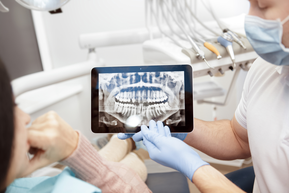
Image Processing
One of the major advantages of digital x-rays is the immediate availability of the images. As soon as the x-ray is taken, the image is transmitted to a computer screen, where it can be viewed and analyzed by your dentist. There is no need for film development, which significantly reduces the waiting time compared to traditional x-rays.
Image Review and Discussion
With the digital image displayed on the computer screen, your dentist can review the results with you right away. Digital x-rays offer high-resolution images that can be zoomed in, enhanced, and adjusted for brightness and contrast. This allows your dentist to provide a detailed explanation of any findings and discuss potential treatment options. Being able to see the images yourself can help you better understand your oral health and make informed decisions about your care.
Storage and Future Access
After the x-ray images have been reviewed, they are stored electronically in your dental records. This digital storage system allows for easy retrieval and sharing of your x-ray images with other dental professionals if needed. It also ensures that your dental history is securely maintained for future reference.
Advancements in Digital X-Ray Technology and Their Impact on Dentists and Patients
Digital x-ray technology continues to evolve, bringing numerous advancements that enhance diagnostic accuracy, patient comfort, and overall dental practice efficiency. These innovations not only improve the quality of dental care but also make the x-ray process more convenient for both dentists and patients. Here are some of the notable advancements in digital x-ray technology and their impacts:
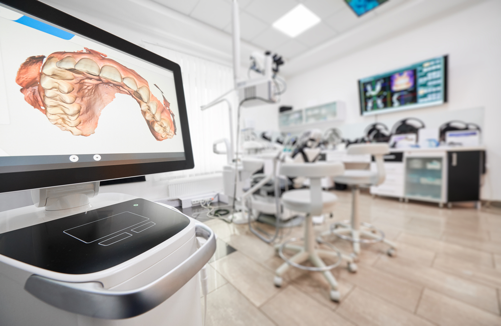
3D Imaging and Cone Beam Computed Tomography (CBCT)
3D imaging and CBCT provide detailed three-dimensional views of the teeth, jawbone, and surrounding structures. This technology offers a comprehensive look at the patient’s oral anatomy, surpassing the capabilities of traditional 2D x-rays.
Impact on Dentists:
- Enhanced Diagnostic Accuracy: Dentists can visualize complex structures more clearly, leading to more accurate diagnoses and treatment plans.
- Improved Treatment Planning: For procedures like dental implants, orthodontics, and oral surgeries, 3D imaging allows for precise planning and execution.
- Increased Confidence: The detailed images reduce the risk of complications during procedures, giving dentists greater confidence in their work.
Impact on Patients:
- Better Outcomes: More accurate diagnostics and treatment planning result in improved treatment outcomes.
- Informed Decisions: Patients can see detailed images of their oral health, helping them understand their conditions and treatment options better.
- Reduced Invasiveness: With precise planning, treatments can be less invasive, leading to quicker recovery times.
Improved Digital Sensors and Phosphor Plate Systems
Modern digital sensors and phosphor plates are smaller, more flexible, and provide higher resolution images with lower radiation doses.
Impact on Dentists:
- Increased Efficiency: High-quality images are captured quickly, streamlining the diagnostic process and reducing appointment times.
- Better Image Quality: Higher resolution images improve the detection of dental issues, allowing for early intervention and more effective treatments.
- Ease of Use: Smaller and more flexible sensors are easier to position, making the x-ray process smoother and more efficient.
Impact on Patients:
- Enhanced Comfort: Smaller sensors are more comfortable to use, reducing discomfort during x-ray procedures.
- Lower Radiation Exposure: Reduced radiation doses make the process safer, particularly for patients requiring frequent x-rays.
- Faster Appointments: Quicker image capture and processing times mean shorter appointments and less time spent in the dental chair.
Cloud-Based Imaging and Data Storage
Cloud-based systems allow for the secure storage and easy access of digital x-ray images and patient records.
Impact on Dentists:
- Streamlined Workflow: Easy access to patient records and images enhances workflow efficiency and reduces administrative burdens.
- Collaboration: Dentists can easily share images with specialists for consultations, improving collaborative care.
- Data Security: Secure cloud storage protects patient data from loss or damage, ensuring records are always available when needed.
Impact on Patients:
- Continuity of Care: Seamless sharing of records ensures continuity of care, even when seeing multiple dental professionals.
- Convenience: Patients can access their records easily, making it simpler to get second opinions or switch providers if necessary.
- Peace of Mind: Secure storage of dental records means patients can trust that their personal information is protected.
Artificial Intelligence (AI) and Image Analysis
AI algorithms can analyze digital x-rays to detect early signs of dental issues, such as cavities, periodontal disease, and oral cancer, with high accuracy.
Impact on Dentists:
- Enhanced Diagnostics: AI tools assist in identifying issues that may be overlooked by the human eye, leading to earlier and more accurate diagnoses.
- Efficiency: Automated analysis saves time, allowing dentists to focus more on patient care rather than image interpretation.
- Standardized Care: AI provides consistent and objective assessments, reducing variability in diagnostic accuracy.
Impact on Patients:
- Early Detection: Early identification of dental problems can prevent more serious issues and reduce the need for extensive treatments.
- Personalized Care: AI-driven insights enable more personalized treatment plans tailored to the specific needs of each patient.
- Confidence in Diagnosis: Patients benefit from the combination of human expertise and AI precision, ensuring comprehensive care.
Conclusion
The advancements in digital x-ray technology are reshaping the landscape of dental care, offering numerous benefits for both dentists and patients. From enhanced diagnostic accuracy and reduced radiation exposure to improved patient comfort and streamlined workflows, these innovations are setting new standards in dental practices. As technology continues to evolve, dental professionals are better equipped to provide precise, efficient, and personalized care. For patients, this means safer, quicker, and more effective treatments, ultimately leading to better oral health outcomes. Embracing digital x-ray technology is a significant step forward in ensuring the highest quality of dental care, making every visit to the dentist a more positive experience.